

Every cell in your body faces a constant challenge: keeping itself healthy. Damaged, misfolded, or excess proteins must be identified and cleared away before they cause harm. Failures in this process are linked to a range of diseases, including neurodegenerative conditions such as Amyotrophic Lateral Sclerosis or ALS and Alzheimer’s disease.
In this new paper from the group, we shed light on a key part of this protein quality control system and reveal, for the first time, the molecular shapes that explain how it works.
Ubiquitin, the cellular “shipping label”
When a cell wants to mark a protein for “shipping”, it attaches a small protein called ubiquitin, a type of molecular “shipping label”. Cells can attach multiple ubiquitin molecules in a chain, and the way these chains are linked together acts as a code that determines where the protein is delivered to.
Two of the most common chain types are linked through a site called K48 or K63. K48-linked chains are shipped to the proteasome for recycling. K63-linked chains in contrast, tend to ship proteins to a different disposal process called autophagy which deals with very large packages, such as larger damaged proteins.
UBQLN2, the cellular “courier”
A protein called UBQLN2 acts as a courier in this system. It recognises the “shipping label”, the polyubiquitin chains, and helps to direct them to the correct disposal site. UBQLN2 has an interesting property as a molecule that allows it to perform this task, known as phase separation.
There are approximately 2-4 million protein molecules per cubic micron in human cells and so there must be processes that allow organisation of this molecular soup. Phase separation is a process where some proteins can spontaneously cluster together to form small, dense droplets inside the cell, akin to the formation of oil droplets when oil is added to water. This process aids in the organisation of proteins in cells and is integral to normal human physiology.
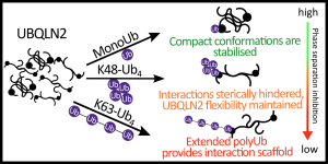
UBQLN2 forms these droplets, and importantly, the type and length of polyubiquitin chain it is bound to has distinguishable impact on the droplets. K48 chains tend to disrupt the droplet formation whereas K63 chains actually promote it.
Until now, the molecular explanations for this difference weren’t understood.
Ion Mobility Mass Spectrometry, how we measured this
To understand what is happening, we needed a technique to look at the 3D shapes that UBQLN2 and polyubiquitin chains adopt when they are interacting. This is very challenging as UBQLN2 is a highly flexible, “disordered”, protein that is less like a rigid Lego block and more like a piece of cooked spaghetti moving around in a pot of boiling water. This means trying to capture the range of shapes it can make, known as the protein conformations, is not possible with many of the gold standard techniques used to study proteins.
Ion-mobility mass spectroscopy (IM-MS) is an excellent technique for studying dynamic protein complexes as the water surrounding the “spaghetti” is evaporated, and the full range of shapes is transferred to be measured. Importantly, there are no biases towards any one type of shape and so the full conformational range can be observed.
What we found
- Chain length controls how open or closed the complex becomes
We looked at UBQLN2 with polyubiquitin chains of increasing length, from a single ubiquitin up to a chain of four. We found that a single ubiquitin is good at forcing UBQLN2 into smaller, more compact shapes and as the chain gets longer, we saw bigger, more elongated shapes. This was very informative and allows us to suggest that the bigger shapes allow interactions to occur that promote formation of UBQLN2 droplets.
- K63-linked chains adopt a surprisingly compact shape
As described before, in an IM-MS measurement, all the water surrounding the protein is evaporated before measurement in the gas phase. When we did this for K63-linked tetraubiquitin, we found it is much more compact than it is in solution. It was also more compact than K48-linked tetraubiquitin. In solution, K63-linked tetraubiquitin has a well known “beads-on-a-string” extended shape where the individual ubiquitin molecules aren’t touching each other. But when all the water is removed, we propose the molecules are all collapsing onto each other as there is nothing keeping them apart. This compaction has not been seen before and allowed us to propose a new molecular mechanism for its behaviour in biology.
- Different shapes explain opposite effects on phase separation
By comparing K48 and K63 tetraubiquitin when bound to UBQLN2, we could begin to explain why the two chain types have opposite effects on phase separation. The K63 chain, even when bound to UBQLN2, retains a propensity to compact and so we think in the complex it isn’t forming interactions with UBQLN2, instead acting as an extendable arm which provides the interactions needed to form the condensates. The K48 chain instead keeps its original properties, and its effect is consistent with a compact species which can disrupt phase separation at high concentrations.
Why it matters
UBQLN2 mutations are associated with inherited forms of ALS, and uncontrolled formation of biomolecular condensates is increasingly recognised as a key feature of neurodegenerative disease. Understanding precisely how ubiquitin chain type and length control UBQLN2’s behaviour is therefore directly relevant to understanding what goes wrong in disease.
More broadly, this work demonstrates the power of ion mobility mass spectrometry for studying protein complexes that are simply too dynamic and heterogeneous for conventional structural biology methods. By revealing the conformational “landscapes” of these we can now connect molecular shape to biological function in a way that wasn’t previously possible.
This is a conceptual step forward in our understanding of how cells use the ubiquitin code to control protein fate.
This work was led by Christina Glen Robb who is a postdoctoral researcher in the lab, in collaboration with the Castaneda lab at Syracuse University, NY and Jakub Ujma at Waters corporation and can be read here: https://pubs.rsc.org/ro/content/articlelanding/2026/sc/d6sc00836d


